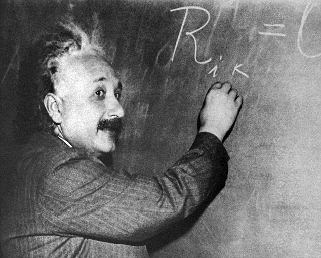
Albert Einstein’s extraordinary genius may have been related to a uniquely shaped brain, a new study suggests.
Researchers compared Albert Einstein’s brain to 85 “normal” human brains to determine, what, if any, unusual features it possessed.
“Although the overall size and asymmetrical shape of Einstein’s brain were normal, the prefrontal, somatosensory, primary motor, parietal, temporal and occipital cortices were extraordinary,” Dean Falk, the Hale G. Smith Professor of Anthropology at Florida State, told Science Daily.
“These may have provided the neurological underpinnings for some of his visuospatial and mathematical abilities, for instance.”
Using 14 recently discovered pictures of the genius’ brain, Dean Falk and her colleagues were able to describe Albert Einstein’s entire cerebral cortex.

Their study, The Cerebral Cortex of Albert Einstein: A Description and Preliminary Analysis of Unpublished Photographs, was published November 16 in Brain, a journal on neurology.
With permission from his family, Albert Einstein’s brain was removed and photographed upon his death in 1955.
It was even sectioned into 240 blocks to make histological slides.
The paper will also outline a “roadmap” to Albert Einstein’s brain made in 1955 by Dr. Thomas Harvey.
Most of those photos, blocks, and slides have been lost from the public eye, and the photographs used by Dean Falk’s team are held by the National Museum of Health and Medicine.